Early screening is the most important and effective step in cancer treatment
Did you know that...
- Almost 50% of dogs over the age of 10 develop cancer.
- Some dog breeds are more prone to cancer than others and are susceptible at a young age.
- Skin tumors represent 1/3 of all tumors in dogs.
Fortunately, less than half of tumors in dogs are cancerous.

Regular check-ups at home
As soon as you notice something suspicious during grooming, such as a lump or bump on or under the skin, make a note of the date, location and size and take a photo.
See your veterinarian as soon as possible, in case it:
- is larger than / equal to one centimeter (pea),
- changes in size, shape or color,
- the fur falls out or the tissue becomes inflamed,
- is present for longer than one month.
Cancer screening with HT Vista®
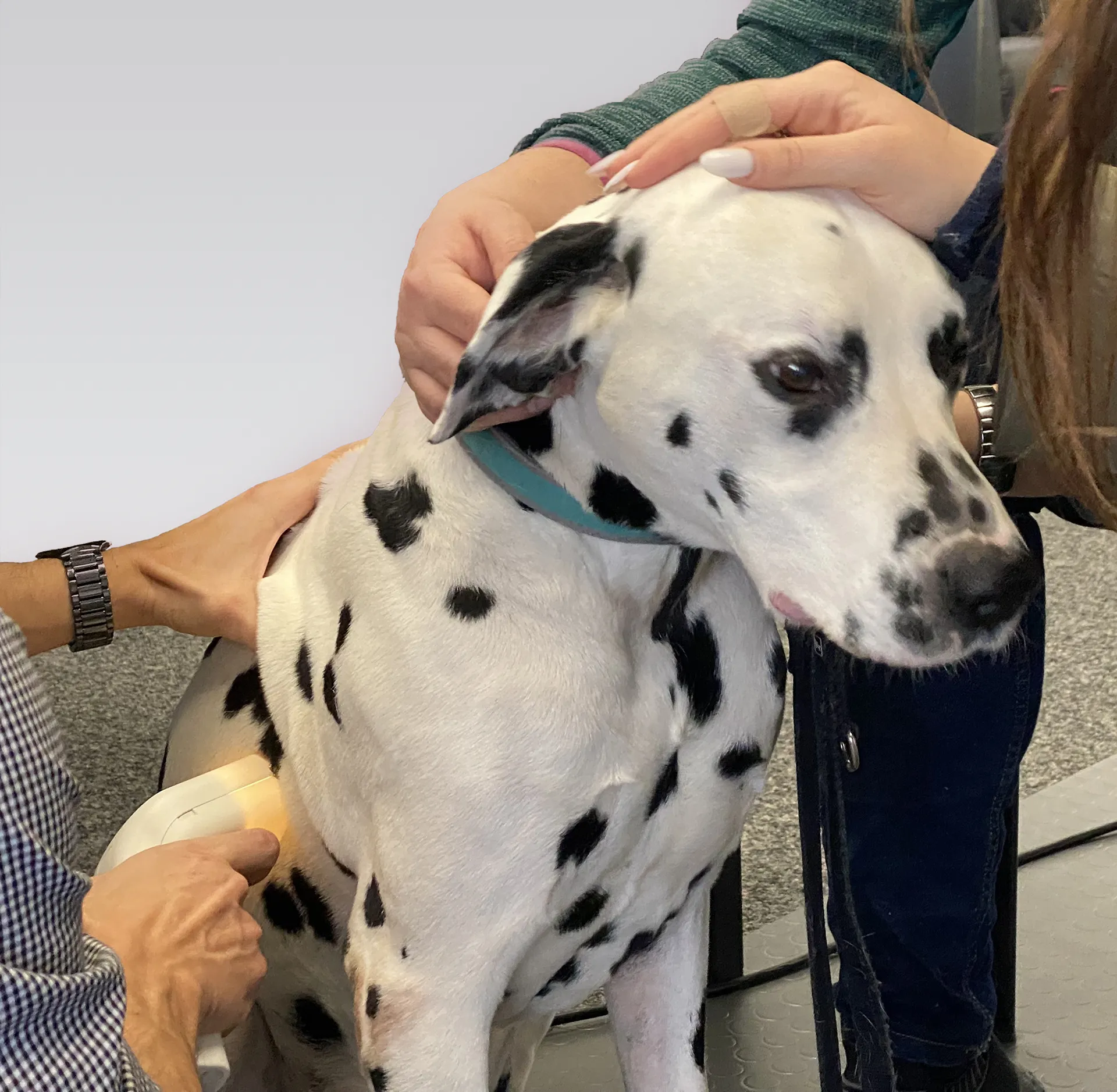
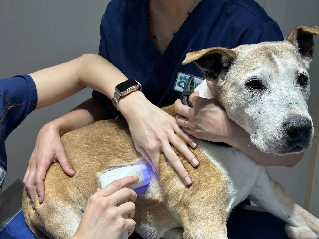
HT Vista® is an innovative system that uses a new patented imaging technology and Artificial Intelligence to simplify the early examination of cutaneous and subdermal masses.
With the non-invasive system, more cases can be painlessly screened directly in the veterinary practice to safely rule out cancer on the spot in a few minutes only!

How does HT Vista® work ?

Registration
Your dog and all lumps and bumps are recorded in the HT Vista system.

Preparation scan
The affected area and a small amount of healthy tissue are shaved.

Scan
The subsequent scan requires 40 seconds of patience and does not disturb your dog.
What are the advantages?

Result in a few minutes
Thanks to artificial intelligence, the tumor tissue is analyzed quickly and you receive the result immediately on site.

Next steps
If there is no negative result, you can discuss the next steps with the vet in good time.

Affordable
No matter how many masses your dog has, screening is affordable and less expensive than other methods.

You will receive the results of the examination in just 5-10 minutes!
A unique new technology
HT Vista® from HTVet is the first AI-driven non-invasive medical device that allows veterinarians and nurses to rule out cancer in subcutaneous and dermal masses thanks to powerful Heat Diffusion Imaging (HDI) technology.
This innovative patented imaging modality relies on unique thermal signals that differ between normal and malignant tissues, as they are recorded by the device, during a rapid heating and cooling process.
Using artificial intelligence, the algorithm compares the patient's signal with previously learned signals and calculates the probability of malignancy.


